Ammonio-Illite-Smect — FTIR1,801 points · 400–4000 cm⁻¹
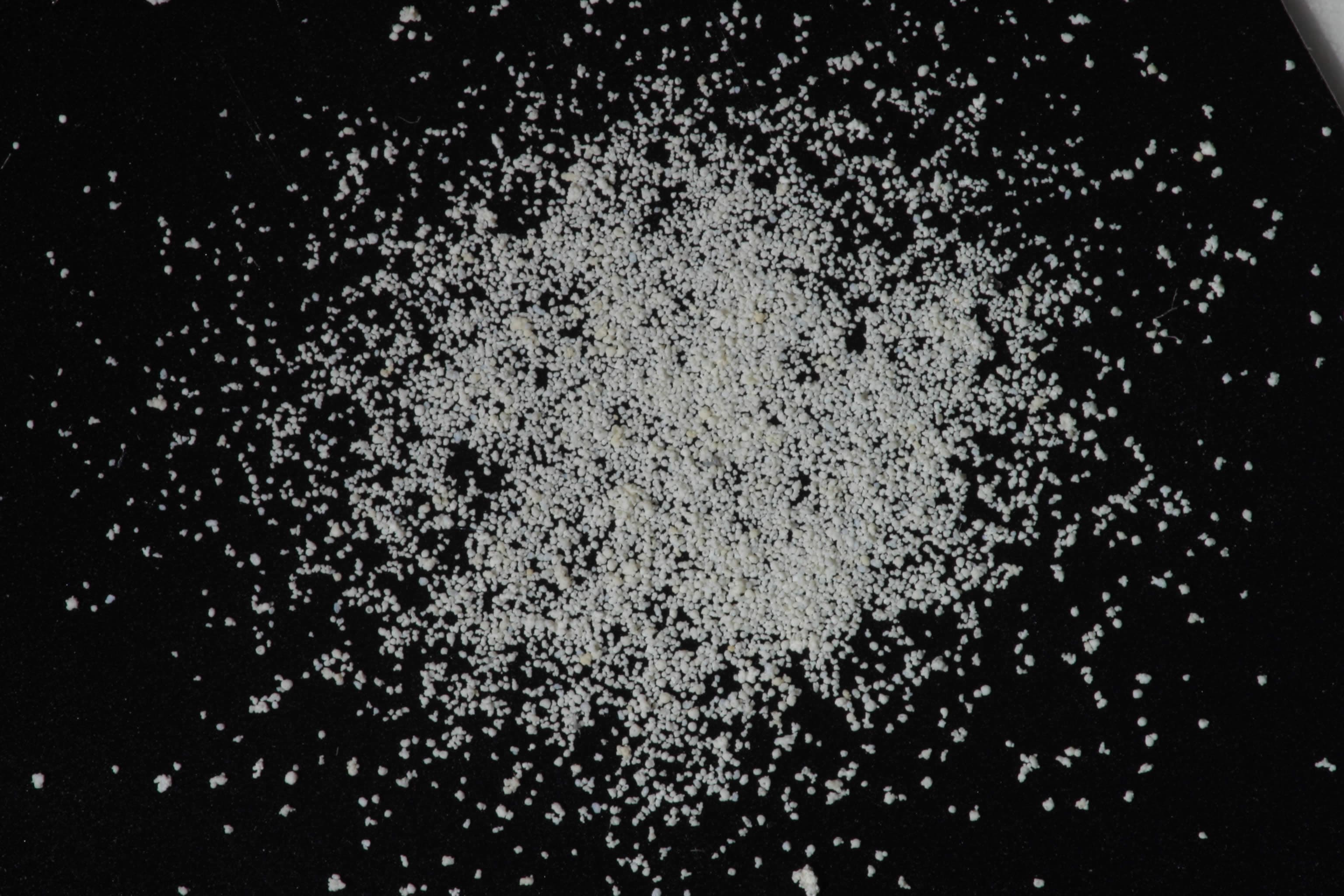
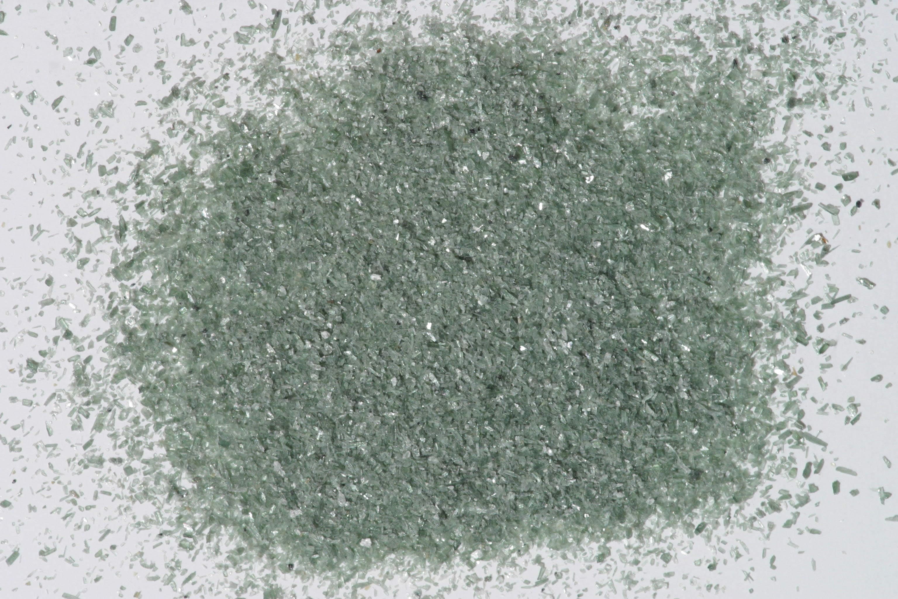
Sample photograph — USGS Spectral Library v7
Spectrum Details
- Modality
- FTIR
- Category
- mineral
- Material Type
- Phyllosilicate
- Sample ID
- GDS87
- Collection Locality
- Synthetic
- Spectral Purity
- 1b2b3b4_ # 1= 0.2-3, 2= 1.5-6, 3= 6-25, 4= 20-150 microns
- Composition / XRD
- None # XRF, EPMA, ICP(Trace), WChem
- Sample Description
- IMAGE_OF_SAMPLE:
- XRD Analysis
- 40 kV - 30 mA, 6.5-9.5 keV File: amilsm87_mdi (smear on quartz plate) References: Deer, Howie and Zussman (1962); Srodon and Eberl (1984); PDF2 #29-1496 Found: Rectorite and/or illite Sought: illite, mascagnite Comment: The pattern consists of broad peaks, some having shoulders or tails extending toward lower d-spacings. The alpha1-alpha2 reflections are not resolved. Both rectorite (29-1495, a 1:1 regularly interstratified dioctahedral mica and dioctahedral smectite) and 1M illite (29-1496) explain most features of the pattern. Neither has a basal reflection exactly in the observed position, 11.0 Angstroms. Weak but sharp and symmetric reflections at 6.10 and 5.45 Angstroms are not identified; in a subsequent bulk mount, the intensity at 5.45 Angstroms was greatly enhanced and that at 6.10 Angstroms greatly reduced. Glycolation of the bulk mount so reduced all intensities that the pattern was useless.
- X Units
- cm⁻¹
- Y Units
- Absorbance
- Data Points
- 1,801
Download
Attribution
USGS Spectral Library Version 7, U.S. Geological Survey
License: Public Domain
DOI: 10.3133/ds1035Analyze in Workbench
Open this spectrum in the SpectralBench workbench for advanced analysis, peak detection, and comparison.
Open Workbench